Case Competition 2023 Case 16 Single-stage treatment of chronic osteomyelitis reconstructed with a free muscle flap
Authors: Josefine Slater, MD, PhD-student, Birgitte Kiil, MD, Alex Ramsden, MD, FRCS (Plast)
A 53-year-old female was referred to the Nuffield Orthopaedic Centre, Bone Infection Unit, Oxford University Hospital, for treatment of a chronic osteomyelitis defect on the left tibia. The patient had a limb laceration leading to a contiguous osteomyelitis of her left tibia that developed at the age of 3 years.
She had previously been treated with a debridement as a child that was left to heal by secondary intention. The chronic wound broke down 2-3 times a year and discharged malodorous pus, but since 2021 it has remained unhealed. The patient is hypothyroid, has a BMI of 35, complete heart block and has a pacemaker since 2014. Antibiotics had been stopped two weeks prior to surgery according to protocol.


Before and after
Patient examination
The patient has a 15x5cm area of scaring on the mid shaft of the left tibia with areas of discharging sinuses from bone. There were palpable tibialis posterior and dorsalis pedis arteries.
Pre-Operative Considerations
Pre-operative planning together with orthopaedic colleagues is paramount. Chronic osteomyelitis is defined as having symptoms for at least six months with clinical and radiological signs in addition to minimum one of the following: the presence of an abscess, a sinus or intra-operative pus, two or more microbiological cultures with the same organisms, or supportive histology[1].
Chronic osteomyelitis has the presence of infected dead bone and biofilm. Antibiotics alone can suppress the infection but not eradicate it. Osteomyelitis may recur if dead bone is not resected, or the soft tissue is not closed over the wound allowing reinfection of bone. The use of local antibiotics is increasingly being advocated as it delivers very high antibiotic doses in the surgical field and nearby tissues [2]. Staged surgical treatment with repeated debridement, and delayed skin closure is common[3]. Also, negative pressure wound therapy has been used, resulting in an increased number of revision surgeries without improving the rate of resolution of osteomyelitis[4]. Single-stage treatment with efficient removal of dead bone and biofilm, sampling of the tissue to elucidate the infecting organism, application of local antibiotics, immediate surgical reconstruction of the bone and soft tissues, followed by systemic antibiotic therapy is the standard treatment, and the procedure planned for, in the presented case[3,5]. Surgical plan for single-stage protocol of osteomyelitis.
– 1 Exposure of bone to allow uncontaminated sampling.
– 2 Multiple deep intra-operative microbiological and histopathological samples following strict protocol.
– 3 Debridement and excision of sinus tracts, scar and dead bone continuing until healthy, bleeding bone and soft tissues are reached.
– 4 Washout with 0.05% aqueous chlorhexidine solution.
– 5 Obturate dead space with local antibiotics biocomposite material.
– 6 Immediate reconstruction of the soft tissues with a free gracilis flap and split-skin graft.
- – Patient information: Risk of flap failure, risk of post-operative fracture, infection recurrence, post-operative infection, wound dehiscence

Step 1: Pre-operative MRI
Pre-operative MRI.

Step 2: Pre-operative MRI
Pre-operative MRI.

Step 3: Incision
Limited exposure to allow for microbiological and histopathological samples.

Step 4: Microbiology sampling
Taking multiple samples for microbiology and histopathology, added to separate tubes and using clean instruments which are changed between each sample to minimise cross contamination. Care is taken to avoid touching the skin or wound with instrument tips, fingers or suction until sampling is complete.

Step 5: Debridement
Debridement and excision of sinus tracts, scar and dead bone continuing until healthy, bleeding bone and soft tissues is reached.

Step 6: Irrigation
Washout with 0.05% aqueous chlorhexidine solution.

Step 7: Deadspace management
Obturation of dead space within bone with antibiotic-loaded biocomposite (Cerament, Bone Support, Lund, Sweden).

Step 8: Flap harvest for immediate reconstruction
Through a longitudinal incision medially on the thigh.

Step 9: Vessel dissection
Dissection of vessels and preparation for microsurgical anastomosis.

Step 10: Recipient vessel dissection
Recipient vessel dissection (a. tibialis post) and preparation. This is often a challenge in the case of chronic infections due to chronically inflamed and fibrotic tissues. Anatomical planes may be disrupted because of previous trauma or surgery.
“The zone of inflammation” may extend far up the limb in the neurovascular plane, why anastomosis often is performed in the scarred area and the vessels are spastic.

Step 11: The gracilis flap
The gracilis flap. Ischemia time was noted. As of Mathes and Nahai classification of pattern of circulation: a type II with dominant pedicle(s) and a minor pedicle (seen with clips). In this patient two minor pedicles and one dominant pedicle was present.

Step 12: Artery anastomosis
End-to-side

Step 13: Vein anastomosis
End-to-end with a 2 mm venous flow coupler.

Step 14: Flap inset.
Flap inset. Tension-free closure with muscle tucked under the edges of the skin defect as it important to ensure a good inset. A drain with weak suction is applied to secure flap to bone surface creating optimal contact, and prevention of fluid collection.

Step 15: Skin graft harvest.
–

Step 16: Final result.
The muscle is resurfaced with a split-thickness skin graft without creation of mesh, but hand fenestrated for a better cosmetic result.
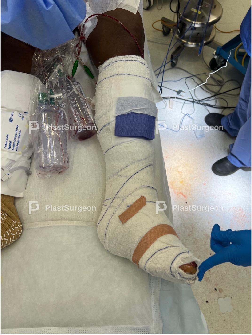
Step 17: Surgical dressing
Surgical dressing.

Step 18: For flap monitoring
–
Step 19: Five day follow-up
Follow-up (5 days) – Flap a bit swollen, but healing as expected.

Step 20: One month follow-up
Follow-up (1 month) – Doing well on antibiotics. – Hyper granulation at the edge of split-skin graft. This is expected to settle down in three to four weeks. – Flap with a good contour.
Post-Operative Plan
– Cast was put on for early post-operative immobilisation.
– Surgical dressing left on constantly for the first five days.
– Create a window to peak for early post-operative monitoring. In this patient, monitoring muscle flap color may be intricate due to the skin pigmentation.
– Flap viability is monitored post-operatively by venous flow doppler. Every 30 minutes until midnight the day of the operation; then hourly for 24 hours; then every other hour for 72 hours; then once per nursing shift until discharge.
– The flow doppler is constantly running, so the patient can alarm the staff if anything changes. – After five post-operative days compress bandaging will be applied reducing edema and improving flap contour. Usually this is worn until no pitting edema, approx. for 2-3 months.
– The patient will then start walking for short periods of time.
– The venous doppler is pulled out at 7-10 days, and the patient was discharged on post-operative day 10.
– The patient will continue oral antibiotic therapy guided by culture results for approx. weeks.
- Single-stage surgery is an effective, patient centric and time efficient treatment.
- Two team operating reduces time in the operating theatre.
- Meticulous tissue sampling with multiple samples
- Compress bandaging for optimal contouring of muscle flaps, reducing the need for corrective surgery later on.
- Failure to sample correctly.
- Failure to excise bone fully
- Recipient vessels are often scarred and spastic.
- The gracillis flap has a short pedicle (approx. 5 cm)
References
- Cierny, G., 3rd; DiPasquale, D. Treatment of chronic infection. J Am Acad Orthop Surg 2006, 14, S105-110, doi:10.5435/00124635-2006
- McNally, M.A.; Ferguson, J.Y.; Lau, A.C.; Diefenbeck, M.; Scarborough, M.; Ramsden, A.J.; Atkins, B.L. Single-stage treatment of chronic osteomyelitis with a new absorbable, gentamicin-loaded, calcium sulphate/hydroxyapatite biocomposite: a prospective series of 100 cases. Bone Joint J 2016, 98-B, 1289-1296, doi:10.1302/0301-620X.98B9.38057.
- Chan, J.K.K.; Ferguson, J.Y.; Scarborough, M.; McNally, M.A.; Ramsden, A.J. Management of Post-Traumatic Osteomyelitis in the Lower Limb: Current State of the Art. Indian J Plast Surg 2019, 52, 62-72, doi:10.1055/s-0039-1687920.
- Diefenbeck, M.; Mennenga, U.; Guckel, P.; Tiemann, A.H.; Muckley, T.; Hofmann, G.O. [Vacuum-assisted closure therapy for the treatment of acute postoperative osteomyelitis]. Z Orthop Unfall 2011, 149, 336-341, doi:10.1055/s-0030-1270952.
- Lorentzen, A.K.; Engel, L.; Gottlieb, H.; Obinah, M.P.B. One-stage treatment of chronic osteomyelitis with an antibiotic-loaded biocomposite and a local or free flap. Eur J Plast Surg 2021, 44, 367-374, doi:10.1007/s00238-020-01754-5.


