Case Competition 2023 Case 10 1st dorsal intermetacarpal artery flap (Foucher flap)
Authors: Andrea Wenger and Volker J. Schmidt, St. Gallen, Switzerland
Initially, the patient presented with a purulent tenosynovitis of the right thumb. Debridement was performed and antibiotics for streptococcus pyogenes administered. 3 weeks later we did another revision surgery because of infected necrose tissue at the pulp of the thumb and osteomyelitis of the terminal bone. After two more debridements this resulted in a soft tissue defect at the palm of the thumb. A gentamycin foam was placed inside the bone and iv antibiotics given although the bacterial cultures were negative.

Before and after
Patient examination
General medical history: 62 year old otherwise healthy male patient with no history of comorbidities (e.g. diabetes), no allergies, non-smoker, no regular intake of any medication Profession: self-employed caretaker, right-handed Right Hand: Skin defect at the palm of the thumb, 2 x 3 cm with exposed bone, FPL tendon, radial and ulnar nerve. EPL and FPL function intact, thumb warm and blood flow intact, 2 point discrimination at the radial side not measurable, 5mm at the ulnar side. X-ray showing signs of osteomyelitis, the IP joint intact
Pre-Operative Considerations
Since the patient was young, healthy, right-handed and a self-employed manual worker, the need of a mechanically robust pulp was the reconstructive goal and amputation at the IP level was no option. 2 flaps would be an appropriate choice for closing this defect: a first dorsal intermetacarpal artery flap or a sensory heterodigital island flap (e.g. ulnar pulp of the ringfinger). Our choice against the latter was due to the history of infection and osteomyelitis with potential risk for further complications and the defect presenting mainly at the radial and therefore the non-dominant part of the pulp in precision grip. The first dorsal intermetacarpal flap (commonly known as Foucher flap) has been initially described by Hilgenfeldt, was designed as a racquet flap by Holevich and modified as an island flap by Foucher. For safety considerations we decided to take a small skin overlying the pedicle and include as many subcutaneous veins as possible in the flap.

Step 1: Flap planning
The surgery is done under plexus block, the patient in a supine position with the shoulder abducted and placed on an arm table. The 1st dorsal intermetacarpal artery is marked by Doppler ultrasound, the flap on the dorsal aspect of the proximal phalanx of the 2nd finger is planned according to the size of the defect. Incisions shouldn’t be extended over mediolateral lines, PIP joint distally and MC joint proximally respectively. The skin overlying the pedicle is included in the flap and the defect later closed primarily.

Step 2: Planning the pivot point
Using a sterile gauze the expected length of the flap and the planned pivot point are again checked before skin incision.

Step 3: Flap rising at the index finger
Inflation of a tourniquet with 250mmHg, surgery is performed using magnifying glasses. Incision of the flap on the index finger and preparation of the subcutaneous tissue leaving the peritendineum of the extensor tendons intact. Include all the subcutaneous veins.

Step 4: Flap rising at the MC level
At the level of the MC joint great care is taken to include the main vessel into the flap. For safety the tourniquet can be released occasionally to check the course of the artery via Doppler ultrasound. Deep arterial branches have to be ligated. Incision of the ulnar border of the skin bridge overlying the 1st dorsal intermetacarpal artery. Preparation is then continued to the pivot point in a deeper layer including the fascia of the 1st dorsal interosseous muscle into the flap.

Step 5: Rising of the flap on to the pivot point
At this point tunnelling of the skin would be possible if the flap were taken as an island flap without a skin bridge. For safety conditions we decided to advance the flap further with the radial border of the skin bridge is incised and preparation of the subcutaneous tissue is continued. Again, great care is taken to include as many veins as possible into the flap.

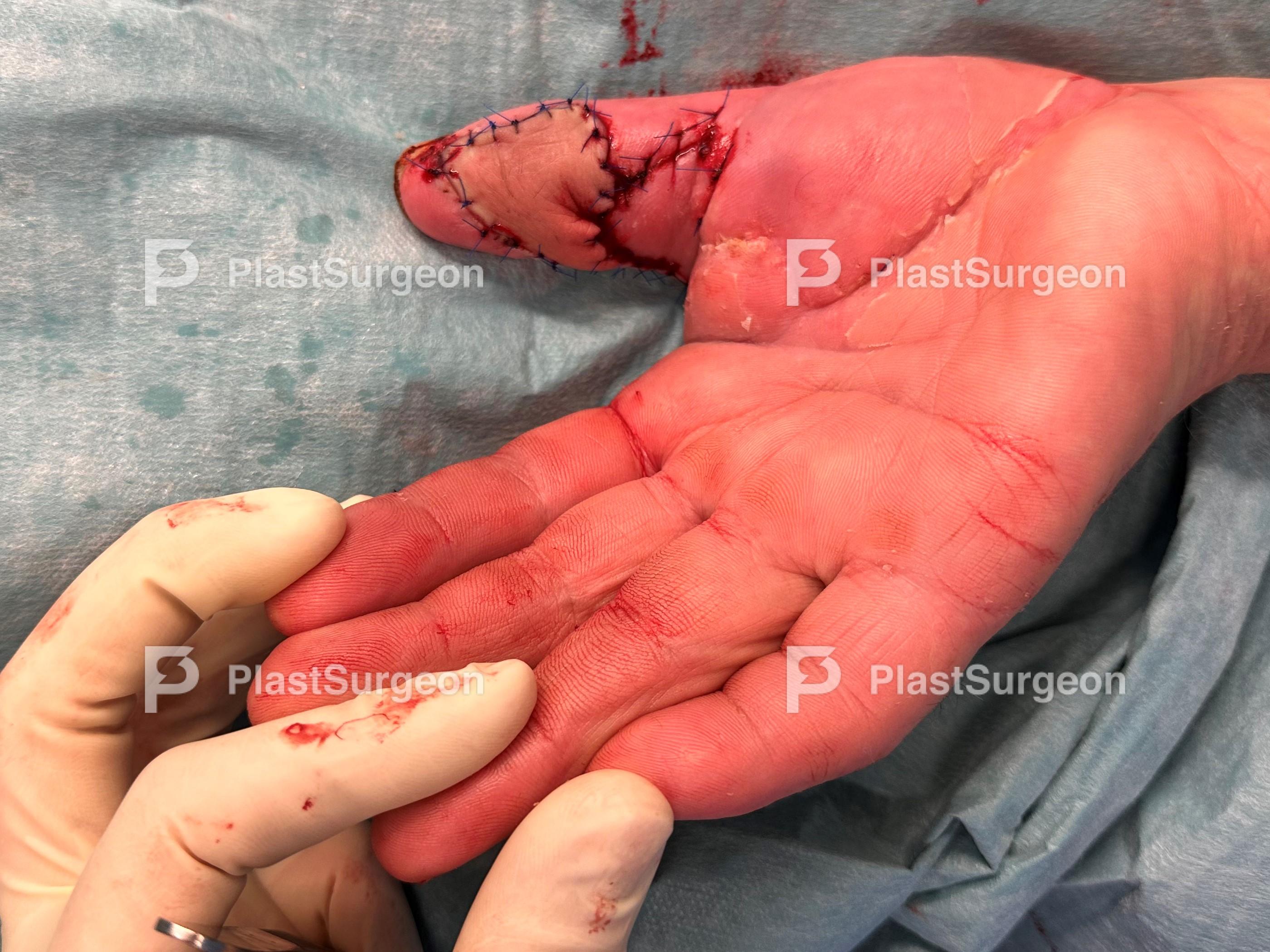
Step 6: Positioning and suturing of the flap, skin grafting the 2nd finger
The flap is then finally rotated from the dorsal aspect of the hand to the pulp of the thumb. In the mediolateral line we did a skin incision to position the flap and sutures are done with 4-0 Prolene. It is crucial to position the flap without tension in full extension and abduction of the thumb so that the maximum width of the fist commissure is preserved.

Step 7: Skin graft the index finger
The skin over the MC joint and metacarpal bone of the index finger is closed primarily. The defect on the dorsal aspect of the 2nd finger is covered with full thickness skin graft from the medial upper arm.
Post-Operative Plan
5 days bedrest, regular flap monitoring every 6 hours, no constrictive dressing Removal of the dressing over the skin graft on the 2nd finger after 5 days and starting with exercises and therapy with focus on the 2nd finger Removal of suture material after 14 days and wearing a compression glove for 3 months, wearing a C-splint for maximum abduction of the thumb during the nights for 3 months in case of contracture of the first web.
- Sensitive flap. Short immobilisation with good range of motion. Regional reconstruction of the tissue. Microsurgical experience is helpful. Preserve paratendineum at the donor site.
- Venous congestion – especially in cases of island flap design. (Partial) flap loss (
References
- Muyldermans T, Hierner R: First dorsal metacarpal artery flap for thumb reconstruction: a retrospective clinical study. Strat Traum Limb Recon 2009;4:27-33
- Couceiro J, Sanmartín M: The Holevic flap revisited: a comparison with the Foucher flap, case series. Hand Surg 2014;19:469-474
- Delokonstantinou IP, Gravvanis AI, et al: Foucher first dorsal metacarpal artery flap versus Littler heterodigital neurovascular flap in resurfacing thumb pulp loss defects. Ann Plast Surg 2001;67:119-122
- Saalabian A, Rab M, et al: Insellappenplastik der ersten dorsalen Mittelhandarterie nach Foucher. Operat Orthop Traumatol 2009;21:614-619
- Kola N: Thumb reconstruction using Foucher’s flap. Opcen Access Macedonian J of Med Sciences 2016;4:70-73
- Tränkle M, Germann G, et al: Neurokutaner Insellappen nach Foucher. Weichteildeckung undn Rekonstruktion der Sensibilität am Daumen. Chirurg 2004;75:996-1002


