Split-thickness skin graft Case 1 Split-skin to lower leg
Authors: Christian Kaare Paaskesen Med. Stud, Eirini Tsigka, MD, MSc and Magnus Avnstorp, MD.
Patient history
75-year-old male with previous excised basal cell carcinoma on the anterior-medial part of lower right crus. The skin was re-excised in a 5 mm margin including subdermal tissue and then covered with a split-thickness skin graft.
Split-skin Procedure

Step 1: Defect is marked
Previous defect is marked in a 5 mm margin.

Step 2: Donor site is shaved
Donor site, medial right thigh, is shaved.

Step 3: Donor area is prepared for grafting
The donor site is disinfected and an area equal to the size of the primary defect is drawn.
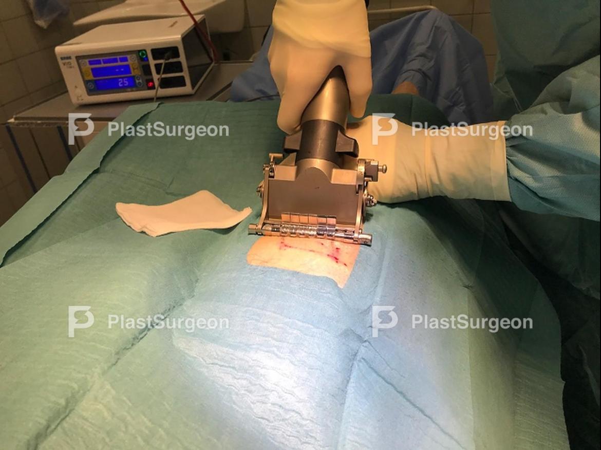
Step 4: Graft is harvested
A skin graft is harvested using an electric dermatome. Donor sites usually heals within 7 days. After harvesting, an adrenalin-soaked gaze is applied, to stop bleeding.

Step 5: Skin is re-excised
The skin is re-excised.

Step 6: Skin is re-excised
The re-excision includes the entire skin down to muscle facia. Make sure sufficient hemostasis has been performed. If blood/fluid is allowed to build up underneath the graft, revascularization can’t happen

Step 7: Graft is meshed
The skin graft is meshed, to prevent blood/fluid to build up and for the graft to cover a larger area. This can also be done using a mesher.

Step 8: Graft is sutured to the wound edges
The graft is sutured to the wound edges, take note of the meshing. The graft should always be sutured in graft to skin direction to better adapt the graft.

Step 9: Dressing is applied
Non-adherent dressing, in the case Nitro- and Jelonet, is applied

Step 10: Bolster is secured
Tie-over dressing/bolster, in this case foam, is applied and sutured to the skin surrounding the defect. The point of this is to create a light pressure on the skin graft, keeping it in place and further preventing hematoma or seroma, without causing necrosis.

Step 11: Donor site is bandaged
Donor site is bandaged.

Step 12: Recipient site is bandaged
Lower leg with the recipient site is further compromised using a compression sock.
References
- Adams DC, Ramsey ML. Grafts in dermatologic surgery: review and update on full- and split-thickness skin grafts, free cartilage grafts, and composite grafts. Dermatol Surg. 2005;31(8 Pt 2):1055-1067. doi:10.1111/j.1524-4725.2005.31831
- Shimizu R, Kishi K. Skin graft. Plast Surg Int. 2012;2012:563493. doi:10.1155/2012/563493
- Thorne, Charles Hm et al, Grabb and Smith’s Plastic Surgery (Wolters Kluwer, 7th ed, 2013;2014;)


