
Loading…
Peer-reviewed case reports, published in our sister journal JPBRS.

Mikkel Halborg Sørensen, Nanja Gotland Sundstrup, Christian Lyngsaa Lang
A 48-year-old woman was referred with a non-radically excised nodular basal cell carcinoma of the anterior scalp complicated by wound infection and dehiscence. Re-excision with 3 mm margins to the galea was performed under local anesthesia. Given local inflammation and uncertain margin status, delayed reconstruction was planned pending histopathological confirmation. After clear margins were verified, controlled subgaleal undermining allowed tension-reduced primary closure. This staged strategy ensured oncologic safety while preserving hair-bearing scalp and avoiding unnecessary flap reconstruction. Patient medical history A 48-year-old woman was referred after incomplete excision of a nodular basal cell carcinoma (nBCC) of the anterior scalp performed in private plastic surgery practice. Histology demonstrated tumor involvement of the lateral margins, while the deep margin was free of tumor. Postoperatively, wound rupture and infection developed. Microbiology showed sparse growth of Staphylococcus aureus, and antibiotic therapy was initiated. The patient was otherwise non-smoking and healthy, with well-treated asthma, as her only relevant comorbidity. Before and After Patient examination Clinical examination revealed a 22 × 19 mm anterior scalp defect with limited perifocal erythema and minimal purulent discharge. No regional lymphadenopathy was palpable. Pre-operative considerations Initial histopathology demonstrated nodular basal cell carcinoma with focal lateral margin involvement and a clear deep margin, without perineural invasion or other high-risk features. In accordance with current recommendations for low-risk BCC, re-excision with 3 mm c
Read at JPBRS
Tushar Dutta, Arun PS, Swati Sattavan
Lumbosacral defects that are too large for local flap reconstruction should be considered for microvascular free tissue transfer. However, lack of suitable recipient vessels in the area makes the task technically difficult. Options such as thoracodorsal and inferior gluteal vessels extended with vein grafts and AV loops have been described. Each option has its own drawbacks. Despite having a short course at its origin, the superior gluteal vessel is a viable alternative. We describe here a case where the vessel was dissected out with adequate length and used as a recipient for free flap reconstruction. Patient medical history The patient was a 41-year-old lady with no known comorbidities. She presented to us with a large lumbosacral mass that had been growing for one year. Before visiting our center, she had been operated twice at other institutions and had recurrences after each occasion. During the last three months the mass had started to grow rapidly and caused ulceration. There was no pain or fever. There was no neurological involvement of lower limbs. Patient had an ECOG status 1. Before and After Patient examination A 17x 22 cm mass was noted encompassing the left part of lumbosacral region and crossing the midline to the opposite side. The superior limit of the lesion was at the L3 vertebra and inferior extent was near the coccyx. Skin ulceration was noted at the center of the mass. On palpation, the mass was warm to touch, hard in consistency and immobile. Skin was fixed and fixity was also noted to the underlying muscle. No tenderness was elicited. Surrounding possible donor sites for flap harvest showed no scars. Pre-operative considerations Preoperative biopsy co
Read at JPBRS
Claes Hannibal Killerich, Nikolaj Warming
A 93-year-old woman presented with a chronic non-healing dorsal finger wound initially diagnosed as actinic keratosis. Repeat biopsy revealed invasive squamous cell carcinoma with joint and bone involvement. Due to the extent of invasion, distal amputation of the third finger was required. Reconstruction was performed using a volar “toilet seat” flap under digital nerve block to minimize surgical burden. The procedure achieved clear margins, preserved stump length, and avoided donor-site morbidity. This case highlights the importance of early re-biopsy of non-healing lesions and adapting surgical strategy to patient age, function, and comorbidity. Patient medical history A 93-year-old woman presented with a wound on the dorsal aspect of the proximal phalanx of the left third finger. A primary biopsy revealed actinic keratosis, and the lesion was treated several times with curettage and electrodesiccation by a private dermatologist. Despite repeated treatments under professional supervision, the wound failed to heal.Due to the persistent non-healing nature of the lesion, a repeat biopsy was performed, confirming the diagnosis of squamous cell carcinoma (SCC). The patient was referred to the Department of Plastic Surgery, Aalborg University Hospital, where a primary excision was performed with a 7 mm surgical margin. Histopathological examination demonstrated invasive tumor growth involving both joint and bone. The defect was subsequently reconstructed using a full-thickness skin graft. Before and After Patient examination The left third finger showed a healed full-thickness skin graft on the dorsal aspect of the proximal phalanx, covering the proximal interphalangeal (PIP) joint. There was no visible residual tumor, and no palpable lymphadenopathy in the left cubital fossa or axillary re
Read at JPBRS
Maria Lerche Mortensen and Philip Månsson
A 62-year-old female presented to the emergency department after a fall from standing position, attempting to break the fall with her left hand. She presented with pain, swelling, and a visible deformity of the wrist. A distal radius fracture of Colles’ type was diagnosed. She was treated in the emergency department in accordance with local guidelines, with closed reduction using both Chinese finger traps and manual reduction with a hematoma block for analgesia. Finally, a dorsal below-elbow cast was applied, and the patient was referred for follow-up after 10-12 days. Patient medical history This patient was active and employed, able to take care of herself, her family and household. She had no past fractures, no relevant medication, and no known allergies. Before and After Patient examination Patient history was obtained, involving circumstances of the trauma, including reasons to suspect underlying disease or other injuries. She fell while walking on a slippery surface, described the trauma as “just bad luck”, she reported no other complaints of pain, had intact memory, and had no cardiac symptoms prior to fall. The clinical examination included inspection, palpation, and function. Inspection: A bayonet deformity was observed, with significant swelling over the radial styloid. There were no wounds or discoloration. Palpation: There was a palpable step-off, and pain over the distal radius. Elbow, hand and fingers were palpated without pain. Therefore, there was no suspicion of other injuries. Function: Neurological and vascular integrity were intact. The motor function was tested: n. ulnaris – finger abduction with intact strength; n. radialis – extension of the MCP-joints; n. medianus – opposition of the first digit. The
Read at JPBRS
Linnea Kristensen Ejiofor
Nail changes can reflect a wide spectrum of benign and malignant conditions, making accurate clinical evaluation essential. This case report reviews key background knowledge on common nail alterations and their differential diagnoses. It aims to support clinicians in recognizing when a nail biopsy is indicated and outlines practical considerations for performing the procedure. In addition, the report describes essential steps in the biopsy technique and provides guidance on appropriate postoperative management and follow-up. Patient medical history A 72-year-old man was referred to the department of plastic surgery and breast surgery at Zealands University Hospital, Roskilde, on suspicion of malignant melanoma under the nail of his right thumb. Before and After Patient examination The patient presented with hutchinson’s sign, longitudinal melanonychia and no swollen lymphnodes. He had no first-degree relatives with melanoma, no prior history of melanoma (invasive or in situ) and no prior history of other skin cancers. Pre-operative considerations Background on Subungual Melanoma (SM) SM is a rare but serious malignancy arising from the nail matrix. Early diagnosis is crucial for treatment and prognosis. Nail changes are often benign; however, malignancy must always be considered in cases of pigmented nail lesions, particularly longitudinal brown or black streaks (longitudinal melanonychia). Biopsy is required when diagnostic uncertainty persists after clinical and dermoscopic evaluation. The incidence of SM is not directly associated with ethnicity in terms of increased risk; however, SM accounts for up to 33% of all melanoma cases in individuals wi
Read at JPBRS
Nanja Gotland Sundstrup, Pia Cajsa Leth Andersen
This case report describes the “Goldilocks procedure” as a technique used for primary breast reconstruction in a patient with prior bilateral c. mamma, treated with lumpectomy and radiotherapy. An implant-based reconstruction was not advised due to the radiotherapy, and she was reluctant to undergo larger scale autologous reconstruction. She was suggested the “goldielocks procedure” as an upgrade to the simple mastectomy which was her alternative consideration. the Goldilocks procedure has the advantage of achieving immediate formation of a small breast, while simultaneously establishing a favorable platform for potential delayed reconstruction with autologous fat grafting. Patient medical history The patient, a 67-year-old woman had a history of bilateral breast cancer: left-sided in 2016 and right-sided in 2023, treated with lumpectomy, and radiotherapy, with additional chemotherapy for the right-sided cancer. Further she was found to carry a pathogenic CHEK2 mutation. The patient underwent bilateral risk reducing mastectomy using the Goldilocks technique, with maximal preservation of the skin envelope and subcutaneous tissue (1). Compared with a simple mastectomy, this approach preserved a good shape and projection resulting in the formation of a small breast. Furthermore, the result provided a more favorable foundation for potential delayed breast reconstruction. The patient was planned for a reconstruction in terms of lipofilling, and mastopexy as well as nipple reconstruction. Before and After Patient examination The patient generally presented with good skin quality. Clinical examination revealed sequelae of prior br
Read at JPBRS
Mikkel Halborg Sørensen, Nanja Gotland Sundstrup, Christian Lyngsaa Lang
A 50-year-old woman with a history of invasive ductal carcinoma (IDC) underwent right-sided papilla reconstruction following secondary deep inferior epigastric perforator (DIEP) flap breast reconstruction. A Tennessee flap technique was used in conjunction with correction of lateral volume of the DIEP flap. The technique provided projection and predictable positioning relative to the contralateral nipple-areola complex. The procedure was performed under local anesthesia in an outpatient setting. The immediate postoperative appearance was satisfactory, and wound healing proceeded without complications. This case higlights the continued utility of the Tennessee flap as a simple, safe, and reproducible technique for delayed nipple reconstruction. Patient medical history A 50-year-old woman with a history of IDC underwent right-sided subcutaneous mastectomy in 2017. In 2025, she underwent unilateral delayed autologous breast reconstruction using a DIEP flap. In 2026, completion of the reconstructive process was planned with papilla reconstruction. The patient was in good general health, non-smoking, and without known comorbidities. Before and After Patient examination Clinical examination demonstrated a well-integrated right-sided DIEP flap, good symmetry compared to the contralateral breast, even though the flap was too voluminous at the lateral side. Absence of nipple-areola complex (NAC) on the reconstructed side and a contralateral areola with soft and not sharply demarcated borders. A well-healed lower abdominal donor-site scar from the DIEP flap harvest was present. Pre-operative considerations In Denmark, the Tennessee
Read at JPBRS
Alaa Jady, Pia Cajsa Leth Andersen
Nipple reconstruction is an important final step in breast reconstruction after mastectomy. This case describes a 42-year-old woman with prior right-sided breast cancer who underwent successful breast reconstruction using a DIEP flap followed by nipple reconstruction with the Tennessee technique. Pre-operative planning focused on achieving symmetry through detailed bilateral measurements and nipple–areola sizing. The procedure involved designing a flap with two arms and a rounded head, careful preservation of the subdermal plexus, and staged suturing. Postoperative follow-up showed good healing, maintained nipple projection, and satisfactory symmetry three months after surgery. Patient medical history A 42-year-old woman with a known history of Hashimoto’s thyroiditis was diagnosed with right-sided breast cancer in June 2022. She received neoadjuvant therapy followed by a right-sided mastectomy in December 2022 and adjuvant radiotherapy. Following completion of these treatments, the patient was deemed cancer-free. In November 2024, she underwent uncomplicated right-sided breast reconstruction using a deep inferior epigastric perforator (DIEP) flap and a contralateral mastopexy for symmetry. Subsequently, right-sided nipple reconstruction was performed in December 2025. Before and After Patient examination Physical examination revealed well-healed scars on both breasts that appeared soft and symmetrical. At the site of the DIEP flap on the right breast, the nipple was absent. Pre-operative considerations Since the patient planned to undergo subsequent nipple–areola complex (NAC) tattooing following our reconstruction, a nipple reconstruction using the “Tennessee” technique was chosen. To achieve symmetry, bilateral measurements were obtained with the patient in the
Read at JPBRS
Lukas Kure-Rosenberg, Magnus Balslev Avnstorp
Background: Proximal dorsal nasal defects are commonly reconstructed with glabellar advancement flaps. However, in selected patients with favorable distal tissue mobility, alternative local strategies may provide equivalent reconstruction while preserving aesthetic subunits thus minimizing scar burden. Case Presentation: An 84-year-old male underwent oncologic excision of a 13-mm moderately differentiated squamous cell carcinoma of the proximal nasal dorsum. The resulting full-thickness defect extended toward the nasal bulb. Preoperative assessment showed significant bulbous skin mobility and distal redundancy without prior surgical scarring. Methods: Instead of glabellar recruitment, reconstruction was performed with a hybrid local advancement strategy combining Peng-inspired medialization with a limited Rintala/H-type advancement component. Tissue was mobilized in the subcutaneous plane to preserve the subdermal plexus, and distal advancement was maintained lateral to the midline to protect dorsal contour symmetry. Results: Flap perfusion remained stable, with no ischemia or venous congestion. At the two-month follow-up, the patient showed a preserved dorsal contour, symmetrical nasal lines, and satisfactory scar placement, without trapdoor deformity or tip distortion. Conclusion: Selected proximal dorsal nasal defects can be reconstructed with hybrid local advancement techniques that strategically exploit distal tissue redundancy. Context-driven modification of established flap principles may allow avoidance of glabellar flaps while maintaining excellent functional and aesthetic outcomes. Patient medical history An 84-year-old male was referred to the Department of Plastic Surgery at Roskilde Sealand University Hospital, Denmark, in winter 2025 by a dermatologi
Read at JPBRS
Ajla Sabitovic, Rikke Børthy Petersen, Nanja Gotland Sundstrup
This case describes the use of a tarsoconjuctival flap for reconstruction of the lower eyelid following excision of a basal cell carcinoma in a 76-year-old woman. Due to the tumor size, depth and location near the limbus of the eye, direct closure was not possible. The tarsoconjuctival flap was chosen as the preferred reconstructive approach because of its ability to reconstruct all layers of the eyelid. Postoperative follow-up at six months demonstrated satisfactory aesthetic and functional outcomes. Patient medical history A 76-year-old woman presented with a biopsy-confirmed nodular basal cell carcinoma involving the right lower eyelid. Tumor excision was performed in general anesthesia with 3 mm margin and intraoperative frozen-section control, confirming tumor free margins. Resection resulted in a full-thickness defect involving approximately 90% of the lower eyelid. Reconstruction was performed with a tarsoconjuctival flap for the posterior lamella and a Tripier flap from the upper eyelid for the anterior lamella. Before and After Patient examination Clinical examination revealed a 12 x 10 mm elevated ulcerating lesion involving the right lower eyelid, extending into conjunctiva and limbus. Pre-operative considerations The patient presented with a full-thickness defect of the lower eyelid involving conjunctiva and the inferior eyelid margin. Due to the tumor size and location direct closure was not possible. Reconstruction with a flap was therefore required to secure reconstruction of all layers of the lower eyelid to give the eyelid full support and thereby secure its function. 1 Step 1 Preoperative skin markings outlining the tumor and 3 mm excision margin. 2 Step 2 Excision of the tumor was performed with a 3 mm margin, involving the conjunctiva, tarsal plate and lower eyelid
Read at JPBRS
Ajla Sabitovic, Nanja Gotland Sundstrup
This case describes the use of full-thickness skin graft for reconstruction of areola in a 64-year-old woman following right-sided mastectomy and delayed breast reconstruction with a DIEP flap. The patient preferred autologous reconstruction and declined tattooing or additional donor-site scars. Due to the light pigmentation on the contralateral areola, it was decided to shave the skin off as a full-thickness in the affected area, and let the following scaring in the area lead to a little change in pigmentation and contration. This approach avoided additional scarring while achieving an acceptable aesthetic result. Patient medical history A 64-year-old woman with a history of right-sided breast cancer underwent mastectomy in November 2022. In August 2024, the patient received a breast reconstruction with a free DIEP flap and a contralateral breast reduction. Nipple reconstruction using af full-thickness skin graft was subsequently performed in March 2025. In October 2025, the patient underwent areola reconstruction with a full-thickness skin graft on the reconstructed breast, combining the donor and recipient site around the nipple. Before and After Patient examination Clinical examination revealed aesthetically pleasing results following the DIEP flap and papil reconstruction as well as a light-colored contralateral areola. Pre-operative considerations It was considered to use a skin-graft from the groin due to its darker pigmentation, but the patient didn’t want any additional scares. Due to the light pigmentation on the contralateral areola, a local full-thickness skin graft was harvested at the recipient site. Postoperative scarring was expected to provide mild pigmentation change and contraction to improve areola definition. 1 Step 1 Preoperative markings performed with the patient in a standing position, showing the neo
Read at JPBRS
RAHUL KAPOOR
Limb salvage in a vessel-depleted extremity remains a formidable reconstructive challenge. We report a case of extensive lower limb gangrene following popliteal artery embolism in a patient with rheumatic heart disease and mitral stenosis. The defect involved exposed bone, necrotic tendons, and absent distal perfusion, with ipsilateral recipient vessels unsuitable for microvascular anastomosis. A cross-leg free flap with external fixation was performed using contralateral posterior tibial vessels. Venous congestion required supercharging with great saphenous vein anastomosis. The postoperative course was uneventful. Flap division at eight weeks demonstrated successful neovascularization, achieving durable limb salvage with progressive sensory and perfusion recovery at 18 months follow-up. Patient medical history Known case of Rheumatic Heart Disease with Mitral Stenosis. Patient developed complain of pain in left leg and foot, in June, 2024. Peripheral Angiography showed 80-90% occluded left external iliac artery & left popliteal artery and 100% left posterior tibial artery. Thrombolysis and plain old balloon Angioplasty done after 3 days of development of pain. Revascularization procedure was followed by development of compartment syndrome in left leg and foot and fever, with progressive discoloration of skin over lower half of leg, ankle and dorsum of foot. Discolouration turned into open wound with complete loss of skin cover, necrosed tendons and exposed bones of lower half of leg, ankle joint and proximal half of dorsum of foot. wound was heavily infected with pus discharge and foul smell. Before and After Patient examination there was progressive discoloration of skin over lower half of leg, ankle and dorsum of foot. Discoloration turned into open wound with complete loss of skin cover, necrosed tendons and e
Read at JPBRS
Abdirahman Abdifatah Mohamed, Numan Omar Ibrahim, Rose Alenyo, Kalanzi Edris
Fibrosarcoma is a rare soft tissue sarcoma with aggressive local infiltration and high recurrence risk. We present a 71-year-old Ugandan man with a giant epigastric fibrosarcoma causing a complex abdominal wall defect. Imaging showed invasion of subcutaneous tissue and rectus muscle without metastasis. The patient underwent wide local excision with 5 cm margins, followed by reconstruction using Prolene mesh and a modified keystone flap with skin grafting. Recovery was uneventful, demonstrating effective surgical and reconstructive management in a resource-limited setting. Patient medical history a 71-year-old male who presented with a two-year history of a progressively enlarging anterior abdominal wall mass. The lesion initially appeared as a small, firm nodule and gradually increased in size, eventually becoming a large, fungating tumor associated with ulceration, necrosis, and intermittent bleeding. There was no history of distant symptoms suggestive of metastasis. Imaging confirmed a locally invasive epigastric mass involving the subcutaneous tissue and rectus abdominis muscle, without visceral or distant spread. Before and After Patient examination On physical examination, the patient had a large, exophytic, fungating mass located in the supraumbilical (epigastric) region of the anterior abdominal wall. The lesion was deeply invasive with irregular margins and was associated with ulceration, areas of necrosis, and active bleeding. The overlying skin was stretched, tense, and partially ulcerated. The mass appeared fixed to the underlying abdominal wall musculature, suggesting muscle involvement. There were no clinical signs of peritoneal invo
Read at JPBRS
Lukas Kure-Rosenberg, Matilda Svenning, Mille Vissing. Magnus Balslev Avnstorp
Background: Reconstructing the full thickness of the nasal ala is technically challenging because it requires restoring contour, lining, and airway patency all at once. The Spear flap, also known as the nasolabial turnover flap, remains a useful single-stage option for selected lateral alar defects. Cases: Two elderly women presented with recurrent basal cell carcinoma of the ala nasi following multiple prior curettages. In case 1, excision resulted in a 20 × 12 mm through-and-through alar defect. In case 2, a recurrent lesion on the left alar measuring 11 × 8 mm initially required staged margin control before final reconstruction. Technique: Both defects were reconstructed using a cheek-based Spear flap after frozen-section-guided excision. Flap design was customized to recreate internal lining and external coverage while minimizing distortion of the alar rim and vestibule. Results: Both flaps remained viable, with no evidence of ischemia or venous congestion. Case 1 achieved complete oncologic clearance and healed with a stable contour; mild residual vestibular fullness did not warrant revision. In case 2, staged clearance allowed for delayed single stage inset, and early healing proceeded without complications. Conclusion: For recurrent through-and-through alar defects, the Spear flap is a reliable and practical reconstructive option. Especially in elderly patients with comorbidities or competing health issues, it provides dependable vascularity, good tissue match, and restores the alar subunit without the complexity of multi-stage procedures. Patient medical history Case 1 (left image) A 90-year-old woman was referred for recurrent basal cell carcinoma of the ala nasi after multiple previous curettages performed in a private dermatology practice in october 2025
Read at JPBRS
Nicolai Lassen Frid, Johan Hindkjær Therchilsen, Jakob Gerlach Christensen
Traumatic partial avulsion of the nasal tip is a rare condition that can present both functional and aesthetic challenges. This case reports a 58-year-old woman with a partial nasal tip avulsion after a fall onto a metal crate. Examination revealed injury to the lower lateral cartilages with loose cartilaginous fragments. Surgical repair focused on restoration of the structural tip support, using end-to-end sutures for approximation of the cartilage and interdomal suturing. This case focuses on rhinoplasty-based principles in acute management of nasal trauma to preserve projection, symmetry, and long-term stability of the nose. Patient medical history A 58-year-old woman presented to the emergency department with a traumatic partial avulsion of the nasal tip sustained after slipping on ice and striking her face against a metal crate. The patient reported neck soreness but exhibited no signs of concussion and no other facial injuries or neck injuries. She had no prior history of nasal trauma. Her medical history included well-controlled hypertension and chronic back pain. Before and After Patient examination Examination of the external nose revealed a large laceration involving the nasal tip, with partial avulsion on the right side. The lower lateral cartilages were injured, with transection of the right lateral crus, a loose fragment of the right intermediate crus, and bilateral fractures of the medial crura with loose cartilaginous fragments. The skin was intact over the columellar and the left alar surface. The wound edges were well defined and with no tissue loss. Sensation was absent in the lacerated tip, but with preserved capillary refill. Anterior rhinoscopy showed a midline septum without h
Read at JPBRS
Sumaira Sattar
A 20-year-old female presented with a severe traumatic injury to the right upper limb following a road traffic accident involving a pedestrian–motorbike collision with subsequent run-over injury. The trauma resulted in extensive soft tissue loss, degloving injury, and associated vascular compromise. Initial management followed advanced trauma protocols, after which the patient was referred for vascular and reconstructive assessment. Imaging revealed brachial artery thrombosis with segmental radial artery injury; however, hand perfusion was maintained through collateral circulation. Following stabilization and vascular reconstruction using a reverse great saphenous vein graft, soft tissue coverage was achieved with an ipsilateral myocutaneous latissimus dorsi flapThe flap provided durable coverage of exposed vital structures and the elbow joint without the need for microsurgical anastomosis. At three-month follow-up, the patient demonstrated excellent functional recovery with satisfactory aesthetic outcome. This case highlights the reliability of the latissimus dorsi flap in complex traumatic upper limb reconstruction. Patient medical history A 20-year-old unmarried female, with no known comorbidities, presented to the emergency department five hours after a road traffic accident involving a bike-versus-pedestrian collision. Following the initial fall, the patient sustained a secondary run-over injury to the right upper limb. There was no history of loss of consciousness, head injury, or trauma to other body regions. Initial assessment and stabilization were performed according to trauma protocols, following which the patient was referred to the plastic and vascular surgery teams for definitive management of the limb injury. Before and After
Read at JPBRS
Ibrahim Güler
A 16-year-old female sustained a crush injury of the left upper extremity, resulting in an open humeral fracture (Gustilo–Anderson IIIC), brachial artery injury, and extensive soft-tissue loss. Initial treatment by trauma and vascular surgery included external fixation and brachial artery reconstruction using a saphenous vein graft. Early graft thrombosis led to critical limb ischemia, and above-elbow amputation was proposed. An emergency limb salvage attempt was performed using a combined free vastus lateralis and anterolateral thigh flap. The limb was successfully salvaged, with a favorable contour and good long-term functional outcome after secondary debulking procedures. Patient medical history A 16-year-old previously healthy female sustained a crush injury to the left upper extremity, resulting in an open humeral fracture (Gustilo–Anderson IIIC), brachial artery injury, and extensive soft-tissue loss [1,2]. Initial management by the trauma and vascular surgery teams at the same hospital included external fixation to stabilize the humeral fracture and reconstruction of the brachial artery using a saphenous vein interposition graft. Early thrombosis of the vein graft led to critical limb ischemia, and above-elbow amputation was subsequently proposed. Before and After Patient examination On examination by the plastic surgery team, the left upper extremity showed extensive soft-tissue loss around the elbow and proximal forearm, with the humerus stabilized in an external fixator. Despite the thrombosis of the brachial artery interposition graft,
Read at JPBRS
Cecilie Jansen, Emil Villiam Holm-Rasmussen, Mette Stueland Wolthers
A 30-year-old previously healthy man was admitted to The Trauma Centre at Copenhagen University Hospital, Rigshospitalet after his left foot was crushed by a 1.5-ton forklift at work. He sustained a large open fracture and an extensive circumferential degloving injury involving the foot and ankle, while distal perfusion remained intact. Computed Tomography (CT) demonstrated multiple intra-articular fractures with suspected Lisfranc injury and no vascular injury on CT angiography. Following orthopedic stabilization, plastic surgical management focused on debridement, tissue viability assessment, skin and heel-pad preservation, and limb salvage. This case highlights the challenges of severe foot degloving and multidisciplinary limb-salvage management. Patient medical history The patient was previously healthy, had no significant past medical history, and was a non-smoker. Before and After Patient examination On arrival at The Trauma Centre at Copenhagen University Hospital, Rigshospitalet, the patient was hemodynamically stable but slightly pale. He reported paresthesia in the left foot, but gross sensation was preserved. Clinical examination revealed an approximately 15 cm open wound laterally on the left foot with exposed bone and tendon. A palpable pulse was present in the dorsalis pedis artery, whereas the posterior tibial artery pulse was not palpable. Capillary refill and sensation were preserved. A trauma CT scan with intravenous contrast, including CT angiography of the lower leg was performed. Imaging demonstrated multiple intra-articular fractures distal to the talus with widening between the second metatarsal and medial cuneiform, raising suspicion of a Lisfranc injury. Air was present in
Read at JPBRS
Mickie Bang Christensen, Magnus Avnstorp
This case presents the use of a modified shark island flap for reconstruction of a defect on the ala nasi following excision of a basal cell carcinoma in a 85-year old male. Due to the tumor size and location, direct closure was not feasible. The shark island flap was chosen as the reconstructive approach. Surgery was done under local anesthesia. Excision of the tumor was done with a 3 mm margin. Histology from frozen sections showed clear margins without malignancy. Postoperative follow up at both one week and two months demonstrated a satisfactory cosmetic outcome and no signs of recurrence. Patient medical history 85-year old male with a medical history of atoxic goiter, hypertension, Parkinson's disease, melanoma and multiple skin carcinomas, was referred with 3 facial skin carcinomas (dorsum nasi, right ala nasi and left side of the upper lip). This case focuses on the carcinoma on the ala nasi. The biopsy showed basal cell carcinoma of nodular subtype. The patient had years prior been operated with a bilobed flap due to a skin carcinoma on the right dorsum nasi, which had resulted in bulging of the right side of the dorsum. Before and After Patient examination Well preserved male with poor skin quality. On clinical examination, a tumor measuring 6 x 5 mm and 2 mm elevation from the surface of the skin was found on the right ala nasi, involving the nasal sulcus. Clinically not adherent to underlying tissue. There were no palpable regional lymph nodes. Pre-operative considerations Surgery and radiation were discussed with the patient. The patient preferred surgery. Due to the tumor size and location, direct closure was not feasible. FTSG would delete the alar-facial sulcus, create a big,
Read at JPBRS
Sondre F. Koren, Jette B. Løvenwald
An 83-year-old male with atrial fibrillation, stage 4 chronic kidney disease, chronic heart failure, monoclonal gammopathy of undetermined significance (MGUS), and glaucoma presented with a 35 × 35 × 15 mm ulcerated squamous cell carcinoma (SCC) of the lateral right lower eyelid. Frozen section-guided full-thickness excision was performed under general anaesthesia. Reconstruction was accomplished in two stages. In the first stage, a combined pedicled tarsoconjunctival flap and a laterally-based periosteal flap (posterior lamella), and a cheek rotation flap (anterior lamella) were used. Frost sutures provided corneal protection during the 4-week inter-stage interval. In the second stage, flap division was performed under general anaesthesia, incorporating Müller's muscle release to prevent upper eyelid retraction. A small dehiscence at the medial flap junction had resulted in a minor notch, which was ultimately resolved with Z-plasty and release of the underlying inferior scar contracture on the cheek. Final histopathology confirmed highly differentiated SCC with clear margins. The multidisciplinary tumour board found no indication for adjuvant therapy. At 23 days following flap division, the patient was satisfied with the functional and aesthetic result. Patient medical history The patient underwent surgery after a 7-week history of a growing tumour of the cheek and lateral lower eyelid. The punch biopsy reported keratoacanthoma/highly differentiated SCC. The tumour had been observed to assess for spontaneous regression, but continued to grow. He had stage 4 chronic kidney disease, treated with Darbepoetin alfa (Aranesp), and glaucoma treated with Latanoprost eye drops, which he had not been taking for several months, with no documented decline
Read at JPBRS
Isha Joshi, Zachary Erlichman, Nia Nikkhahmanesh, Christina Chopra, Jason Ganz
Background: Tissue from non-salvageable limbs may be used in reconstruction of complex defects and represent the principle of the “spare- parts” concept.[1] Fillet flaps advantages include soft tissue coverage of large defects, biocompatibility of an autologous donor, and donor tissue resembling native tissue due to proximity. Case: Fewer than 100 hemicorporectomies have been published in the literature.[2] We present the case of a 53-year-old male who underwent hemicorporectomy for recurrent gluteal sarcoma followed by reconstruction using a near-total thigh fillet flap. Conclusions: Fillet flaps provide soft-tissue coverage of large defects, biocompatible autologous tissue, and donor tissue resembling native anatomy. Patient medical history Primary Diagnosis: In 2015, the patient was diagnosed with a 15-cm high-grade (grade 3) spindle cell carcinoma thought to represent a fibrosarcoma with transformation to dermatofibrosarcoma protuberans, located superior to the buttock. The tumor was resected at an outside hospital (OSH) without perioperative complications. First Recurrence: In 2016, the patient presented to the OSH with recurrent mass in the tumor bed measuring 12.2 cm with erosion of lower coccyx, extension into posterior pelvis, and deviation of the anus. He underwent neoadjuvant radiation and systemic therapy. In 2017, he underwent tumor debulking and permanent diverting colostomy placement. After relocating to New York, he established care at our institution and subsequently underwent conversion of a loop colostomy to an end stoma with mucous fistula in 2020. Secondary Recurrence: In 2022, imaging demonstrated a recurrent tumor above the buttock. The pat
Read at JPBRS
Miss Ketki Kaushal, Mr Oliver Bassett
Gorlin-Goltz syndrome, also known as Nevoid Basal Cell Carcinoma Syndrome, is a rare autosomal dominant condition associated with pathogenic variants in the PTCH1 gene and dysregulation of the Hedgehog signaling pathway. A patient with longstanding disease presented with recurrent periocular basal cell carcinoma requiring Mohs excision, resulting in a large full-thickness lower eyelid defect. Reconstruction was performed using a hard palate graft for posterior lamella replacement and a radial forearm free flap with palmaris longus tendon sling for anterior lamella and structural support. Microvascular anastomosis to facial vessels was performed. This case highlights the reconstructive challenges of periocular defects in NBCCS and emphasises multidisciplinary management. Patient medical history A 59-year-old male with a longstanding history of Gorlin–Goltz syndrome (Nevoid Basal Cell Carcinoma Syndrome) diagnosed in 1997 has been under multidisciplinary care involving dermatology, plastic surgery, oncology, and ophthalmology for multiple recurrent basal cell carcinomas. His medical history includes hypertension, iron deficiency anaemia secondary to diverticulosis, chronic gastritis with duodenal ulcer disease, and melanoma in situ of the right knee excised in 2016. He has undergone numerous treatments for cutaneous malignancies including repeated surgical excisions, Mohs micrographic surgery, topical therapies (5-fluorouracil and imiquimod), and reconstructive procedures. In 2012 he underwent Mohs surgery for an infiltrative basal cell carcinoma of the left lower eyelid. Over subsequent years, multiple lesions were treated across the face, temples, ears, forearm, and calf. Since April 2016 he has been managed with the He
Read at JPBRS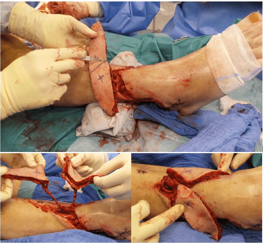
Luisa Lotter¹, Marta Jezierska¹, Ilja W. Käch¹, Volker J. Schmidt¹
46-year-old man presented with two full-thickness soft-tissue defects of the distal right lower leg (medial 7 × 4 cm, lateral 10 × 5 cm) with exposed tendons, osteosynthesis material, and bone after a Gustilo grade IIIb open tibial and fibular fracture. During initial orthopedic stabilization, interdisciplinary orthoplastic assessment and planning were performed. Because of exposed functional structures and two spatially separated defects with healthy anterior skin between them, microsurgical free-tissue reconstruction was chosen to provide stable coverage while preserving the anterior skin and preventing flap-related functional impairment. Reconstruction was achieved using a split anterolateral thigh (ALT) flap based on two independent perforators, enabling simultaneous coverage of both defects with a single vascular pedicle. Patient medical history The patient’s medical history was significant for type 2 diabetes mellitus, two-vessel coronary artery disease, and obesity. Before and After Patient examination Clinical assessment revealed two full-thickness defects at the distal lower leg: • medial defect: 7 × 4 cm with exposed tendons • lateral defect: 10 × 5 cm with exposed bone and osteosynthesis material Both wounds showed non-viable soft-tissue components requiring radical surgical debridement prior to reconstruction. An CT-angiography was performed, revealing a regular three-vessel supply to the affected lower right leg. Preoperative color-duplex-ultrasound was routinely performed to identify the ALT perforator pattern and to verify, if a two-perforator split ALT-fashion is possible. Due to our tertiary orthoplastic program final interdisciplinary reconstruction was schedule
Read at JPBRS
Nelson Ramirez Lozano
We present the successful case of a patient who sustained severe trauma to the upper extremity caused by a sugarcane mill (trapiche). We consider this type of trauma to be a reconstructive challenge given the significant degree of soft tissue compromise, the presence of infection in nearly all such cases, and the imperative need for these patients to return to their daily activities. A propeller flap based on two perforators of the scapular artery was designed and executed, enabling adequate reconstruction and wound coverage without generating donor-site morbidity, facilitating early rehabilitation and rapid functional recovery. Patient medical history A male patient sustained severe trauma to the left upper extremity involving a sugarcane mill during occupational activities. He was admitted to the emergency department, where hemostasis was achieved and hemodynamic stabilization was attained. On initial evaluation, no fractures were identified; however, the patient presented with humeral bone exposure, circumferential soft tissue loss involving the upper arm, restricted range of motion, and altered sensation. He subsequently underwent multiple irrigation and debridement procedures, as well as serial negative pressure wound therapy (NPWT) dressing changes performed by the Orthopedics and Traumatology service, achieving adequate control of necrotic tissue and resolution of infection. Plastic Surgery consultation was then requested. Before and After Patient examination The patient presented with a circumferential soft tissue defect of the left upper arm with exposed humerus, no active signs of infection, and an in-situ negative pressure wound therapy device. Physical examination a
Read at JPBRS