DIEP Flap Procedure
Authors: Rami Mossad Ibrahim, Magnus Obinah, Magnus Balslev Avnstorp MD, Peter Stemann and Birgitte Jul Kiil
Procedure (Photos)
1
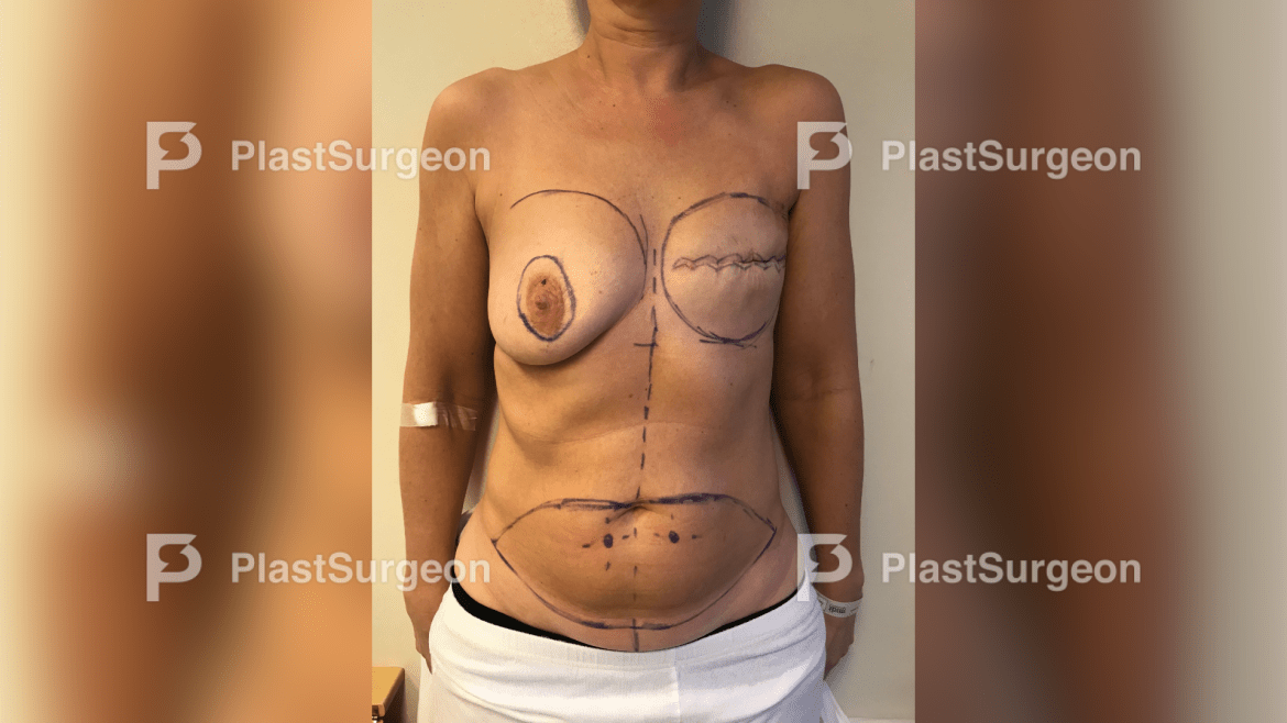
Step 1: Pre-operative drawing
Pre-operative drawing of donor site with two large periumbilical arterial perforators (a DIEP flap) and recipient site (internal mammary artery of left breast)

2
Step 2: Harvesting of perforator flap

3
Step 3: Microsurgical anastomosis
Microsurgical anastomosis of vessels on recipient site using clips

4
Step 4: Closure of abdominal donor site/defect.

5
Step 5: Suturing of DIEP flap to the left breast
Suturing of DIEP flap to the left breast (recipient site) – The reconstructed breast
Instruction to the patient
Pre-operative regimen
Post-operative calm regime.
Follow-up
Clinical flap assessment by laser and/or hand ultrasound doppler every hour the first 24 hours and the following days.
See: **LINK TO CHAPTER ON MONITORING MICROSURGERY FLAPS**
Depending on the complexity of the flap the monitoring may be less extensive.
Pearls and Pitfalls when performing perforator flaps
- Overall use Diligence (careful and persistent work) and intelligence.
- Use appropriate imaging technology if possible.
- Make sure that you have a recipient vessel in mind when planning a free perforator flap.
- Have a Plan A, Plan B, Plan C if the vessel is no good.
- Be aware and respectful that harvesting a perforator flap is difficult.
- Remember that small perforator flap vessels can be anastomosed to other small perforator vessel as recipient vessels, which can be useful on legs etc.
- Practice using magnifying glasses (loup glasses).
- In case you have a small defect ie on a leg following cancer resection, then practice looking for a perforator flap nearby. Try to do a propeller flap instead of a random flap or a skin transplant.
- Minor perforator flaps are also very useful in the face.
- Look after perforator vessels near the defect with ultra sound before the resection.
- The perforator vessels are often very small and fragile. Be careful and have fun!
References
- Koshima I, Soeda S. Inferior epigastric artery skin flaps without abdominis muscle 1989:645–8.
- Blondeel PN, Van Landuyt KH, Monstrey SJ et al. “the ”gent“ consensus on perforator flap terminology: preliminary definitions”. plast. reconstr. surg. 112 (5): 1378–83; quiz 1383, 1516; discussion 1384–7. 2003.
- Color Doppler ultrasonography targeted reconstruction using pedicled perforator flaps—a systematic review and meta- analysis- Rami Mossad Ibrahim, Gudjon Gunnarson, Javed Akram, Jørn Bo Thomsen and Jens Ahm Sørensen.


